|
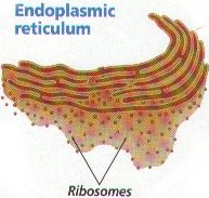
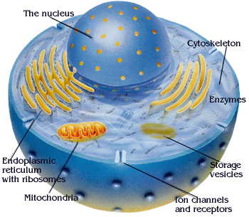
The endoplasmic reticulum is a system of fluid-filled canals,
or channels , enclosed by membranes. These canals usually form a continuous network throughout the cytoplasm. The canals
of the endoplasmic reticulum serve as paths for the transport of materials through the cell. eg proteins.
Rough endoplasmic reticulum bears the ribosomes during protein synthesis. The newly synthesized
proteins are sequestered in sacs, called cisternae . The system then sends the proteins via small vesicles to the Golgi Complex , or, in the case of membrane proteins, it inserts them into the membrane. Rough endoplasmic reticulum may either be vesicular or tubular.
Or it may consist of stacks of flattened cisternae (like sheets) that may have bridging areas connecting the individual sheets.
The Ribosomes sit on the outer surfaces of the sacs (or cisternae). They resemble small beads sitting in rosettes or
in a linear pattern.
Smooth endoplasmic reticulum is found in a variety of cell types and it serves different functions
in each. It consists of tubules and vesicles that branch forming a network. In some cells there are dilated areas like the
sacs of rough endoplasmic reticulum. The network of smooth endoplasmic reticulum allows increased surface area for the action
or storage of key enzymes and the products of these enzymes. In the case of smooth endoplasmic reticulum in muscle cells,
the vesicles and tubules serve as a store of calcium which is released as one step in the contraction process. Calcium pumps
serve to move the calcium.
f.
Chloroplast!!
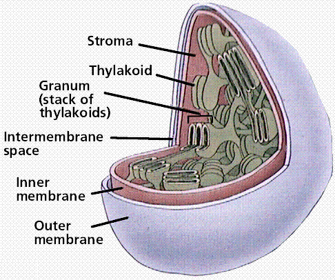
Chloroplasts are the green colored pigment and they
help in the following process. The main reaction that takes place in chloroplasts is known as the process of Photosynthesis.
In this process carbondioxide, water and the energy obtained from sunlight react together to form glucose and oxgen.
Chloroplasts are found in plant cells only and in protists. The Many Reactions that occur during the photosynthesis
in plants can be grouped into two broad categories.
1. Light reactions (photosynthetic reactions): in this
step, energy derived from light energizes an electron in the green organic pigment chlorophyll, enabling electron to move
along an electron-transport chain in the thylakoid membrane. The chlorophyll obtains its electrons from water by producing
oxygen as a by products. During the electron transport process H+ is pumped across the thylakoid membrane, and resulting electrochemical
proton gradient drives the synthesis of ATP in the stroma. In final step high energy loaded onto NADP+.
2. Dark reactions (carbon fixation reactions). ATP and
NADPH that are produced in first step are going to be utilized for fixing CO2 into sugar.
The Calvin Cycle :
The cycle spends ATP as an energy source and consumes NADPH2 as reducing power for adding
high energy electrons to make the sugar. There are three phases of the cycle. In phase 1 (Carbon Fixation), CO2
is incorporated into a five-carbon sugar named ribulose bisphosphate (RuBP). The enzyme which catalyzes this first step is
RuBP carboxylase or rubisco. It is the most abundant protein in chloroplasts and probably the most abundant protein on Earth.
The product of the reaction is a six-carbon intermediate which immediately splits in half to form two molecules of 3-phosphoglycerate.
In phase 2 ( Reduction), ATP and NADPH2 from the light reactions are used to convert 3-phosphoglycerate to glyceraldehyde
3-phosphate, the three-carbon carbohydrate precursor to glucose and other sugars. In phase 3 (Regeneration), more ATP is used
to convert some of the of the pool of glyceraldehyde 3-phosphate back to RuBP, the acceptor for CO2, thereby completing
the cycle. For every three molecules of CO2 that enter the cycle, the net output is one molecule of glyceraldehyde
3-phosphate (G3P). For each G3P synthesized, the cycle spends nine molecules of ATP and six molecules of NADPH2.
The light reactions sustain the Calvin cycle by regenerating the ATP and NADPH2.
The Calvin Cycle
The cycle spends ATP as an energy source and consumes NADPH2 as reducing power for adding high energy electrons
to make the sugar. There are three phases of the cycle. In phase 1 (Carbon Fixation), CO2 is incorporated into
a five-carbon sugar named ribulose bisphosphate (RuBP). The enzyme which catalyzes this first step is RuBP carboxylase or
rubisco. It is the most abundant protein in chloroplasts and probably the most abundant protein on Earth. The product of the
reaction is a six-carbon intermediate which immediately splits in half to form two molecules of 3-phosphoglycerate. In phase
2 ( Reduction), ATP and NADPH2 from the light reactions are used to convert 3-phosphoglycerate to glyceraldehyde
3-phosphate, the three-carbon carbohydrate precursor to glucose and other sugars. In phase 3 (Regeneration), more ATP is used
to convert some of the of the pool of glyceraldehyde 3-phosphate back to RuBP, the acceptor for CO2, thereby completing
the cycle. For every three molecules of CO2 that enter the cycle, the net output is one molecule of glyceraldehyde
3-phosphate (G3P). For each G3P synthesized, the cycle spends nine molecules of ATP and six molecules of NADPH2.
The light reactions sustain the Calvin cycle by regenerating the ATP and NADPH2.
PHOTOSYNTHESIS CHEMICAL REACTION:
CO2 + H2O + Energy = C6H12O6 + O2
(carbon
dioxide) (water) (sunlight) (glucose) (oxygen)
what is the role of mitochondria in making stored chemical-bond energy available to cells by
completing the breakdownof glucoseto carbon dioxide?
Mitochondria
g.
The chemical energy generated by chloroplasts is stored in the bonds of sugar molecules untill they
are broken down by mitochondria. Mitochondria are memebrane-bound organelles in plant and animal cells that transform energy
for the cell. This energy is then in the bonds of other molecules that cell organelles can access easily and quickly when
energy is needed. The energy for storing these molecules is obtained from completing the breaking down of glucose to
carbon dioxide. The main job of mitochondria is the cellular respiration.It reacts totally opposite to that in chloroplast.
It combines with glucose and oxygen to give carbon dioxide, water and energy in the ATP ( adenosine triphosphate ) form. C6H12O6 + O2 = CO2 + H2O + Energy .
Mitochondria are usually found in all eukaryotic cell eg. protists, fungi, animal cell and in plant cells.
Cellular respiration is the process of oxidizing food molecules, like glucose, to carbon dioxide and
water. The energy released is trapped in the form of ATP for use by all the energy-consuming activities of the cell.
The process occurs in two phases:
- glycolysis, the breakdown of glucose to pyruvic acid
- the complete oxidation of pyruvic acid to carbon dioxide and water
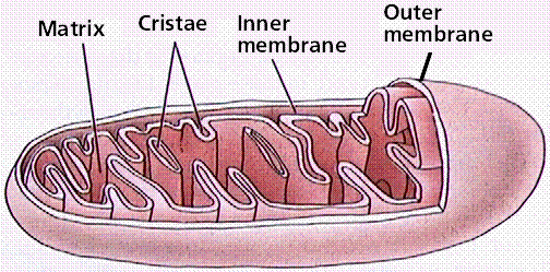
Mitochondria are membrane-enclosed organelles distributed through the cytosol of most eukaryotic cells. Their main function is the conversion of the
potential energy of food molecules into ATP.  Mitochondria have: Mitochondria have:
- an outer membrane that encloses the entire structure
- an inner membrane that encloses a fluid-filled matrix
- between the two is the intermembrane space
- the inner membrane is elaborately folded with shelflike cristae projecting into the matrix.
- a small number (some 5–10) circular molecules of DNA
- This 2-carbon fragment is donated to a molecule of oxaloacetic acid.
- The resulting molecule of citric acid (which gives its name to the process) undergoes the series
of enzymatic steps shown in the diagram.
- The final step regenerates a molecule of oxaloacetic acid and the cycle is ready to turn again.
Each of the 3 carbon atoms present in the pyruvate that entered the mitochondrion leaves as a molecule
of carbon dioxide (CO2).
At 4 steps, a pair of electrons (2e-) is removed
and transferred to NAD+ reducing it to NADH + H+.
At one step, a pair of electrons is removed from succinic
acid and reduces FAD to FADH2.
The electrons of NADH and FADH2 are transferred to the respiratory chain.
CYTOSOL : The
cytosol is the "soup" within which all the other cell organelles reside and where most of the cellular metabolism occurs.
Though mostly water, the cytosol is full of proteins that control cell metabolism including signal transduction pathways,
glycolysis, intracellular receptors, and transcription factors. Cytoplasm
is a collective term for the cytosol plus the organelles suspended within
the cytosol.
Oxidation of glucose is known as glycolysis.Glucose is oxidized to either
lactate or pyruvate.
Cristae are the infoldings of the inner membrane of a mitochondrion. They are studded with proteins, including ATP synthase and a variety of cytochromes, and function in cellular respiration. They provide more surface area for cellular respiration to happen.
- Protons are translocated across the membrane, from the matrix to the intermembrane space
- Electrons are transported along the membrane, through a series of protein carriers
- Oxygen is the terminal electron acceptor, combining with electrons and H+ ions to
produce water
- As NADH delivers more H+ and electrons into the ETS, the proton gradient increases,
with H+ building up outside the inner mitochondrial membrane, and OH- inside the membrane.
A connection between d,e,f and g
Firts of all, all teh substances described in d,e,f and g are all teh members of a cell. In all
the descriptions, these membranes have been described very minutely along with their roles and funcitons, which they carry
out. Proteins is the main connection between all these points. Bullet d refers to the path of transfer for proteins to the
cytoplasm with the help of ribosomes and RNA. Bullet e refers to the help provided by the endoplasmic reticulum and golgi
bodies in the secretion of proteins to the nucleus. Bullet f indirectly states how protein is obtained by chloroplasts. In
this process, chloroplasts capture the energy form the sunlight, which is a protein to them for their sustainance. Bullet
g too indirectly states how protein is provided to other cells by mitochondria. Mitochondria is known as, "The Powerhouse
of Energy". It stores the chemical bond energy within itself and when needed provides this energy(proteins) to other cells
h. types of macromolecules
Polysacchrides
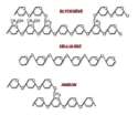
Polysaccharides (sometimes called glycans) are relatively
complex carbohydrates. They are polymers made up of many monosacchrides joined together by glycosidic linkages. They are therefore very large, often branched,
molecules. They tend to be amorphous, insolubile in water, and have no sweet taste. When all the constituent monosaccharides
are of the same type they are termed homopolysaccharides; when more than one type of monosaccharide is present they
are termed heteropolysaccharides.
Examples include storage polysaccharides such as starch and glycogen and structural polysaccharides such as cellulose
Proteins
proteins are compounds that contain nitrogen as well as carbon, hydrogen, and oxygen. Some proteins
also contain sulfur and phosphorus. The number of possible proteins is virtually unlimited. Proteins have an astonishing range
of properties. Proteins make life possible in its present degree of complexity. There are proteins in the structural parts
of cells and body tissues, such as cartilage, bones, and muscles. There are proteins in the hormones, the chemical messengers
that regulate body functions in plants and animals. There are proteins in antibodies, the substance that protects animals
against disease organisms.
The Nucleus and the Nucleic Acids: RNA and DNA
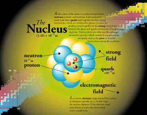
Nucliec acids are compounds that contain phosphorous and nitrogen in addition to carbon,hydrogen,and
oxygen. There are two kinds of nucleic acids. One is called DNA and RNA. These substances were first found in the nucleus.
DNA is the heriditary material that is passed on from one generation to the next during reproduction.
A nucleotide is a monomer or the structural unit of nucleotide chains forming nucleic acids as RNA and DNA. A nucleotide consists of a heterocyclic nucleobase, a pentose sugar (ribose or deoxiribose), and a phosphate or polyphosphate group. Nucleotides also play important roles in cellular energy transport and transformations (notably ATP and NAD+/NADH), and in enzyme regulation (see for example, protein kinase).
PRECURSOR: In biological processes especially
metabolism, a precursor is a substance from which another, usually more active or mature
substance is formed.
For instance, certain liver enzymes are precursors to insulin. Or, this is the opposite of the body breaking down a more complex substance, like protein into individual chemicals for use within the body.
Lipids
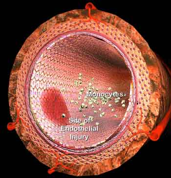
Lipids are a part of cell structures and serve as a reserve energy supply in an organism. Lipids
furnish about twice as much energy as the same ammount of carbohydrates. They include the substances commonly called fats,
oils and waxes. Like carbohydrates, lipids are made of crabon,hydrogen and oxygen. However there is less oxygen in lipids
than in carbohydrates. Fats and oils are chemically similar.Unlike fats, however, oils remain liquid at room temperature.
Plants store oils in seeds. Some familiar oils are peanut oil, corn oil, and castor oil.
Polysacchrides
|
+
cellulose
|
-
Fructose
|
|
chitin
|
glucose
|
|
starch
|
galactose
|
|
glycogen
|
maltose
|
|
animal starch
|
sucrose
|
nucleic acids
+
Ribo Nucliec Acid (RNA)
|
|
Deoxyribose Nucleic Acid (DNA)
|
|
|
oxygen
|
guanine
|
|
hydrogen
|
cytosine
|
|
phosphorus
|
uracil
|
protiens
lipids
j
How does cytoskeleton and cell wall give shape and internal organisation to a eukaryotic cell?
Eukaryotic cells make up organisims from the Protist, Fungi, Plant, and Animal Kingdoms
(That's Mostly All). Eukaryotic cells have large diameters, 10 micrometers and up, which is large in comparison to Prokaryotic
cells which range from 5 through 2 micrometers. The size of Eukaryotic cells is small compared to the size of humans but is
large in the microbial world.
The largest difference of a Eukaryotic cell from a Prokaryotic cell other than size is
the presence of a Nucleus. A Nuclus is a membrane which protects DNA against the elements of the cell. This "Nuclear Envelope"
lets Transfer RNA through and keeps ribosomes out. Ribosomes change Transfer RNA into proteins which perform functions in
the cell.
Other important objects in a Eukaryotic cell are Mitochondria. Mitochondria produce energy through the burning
of Glucose, a sugar. These mitochondria are roughly the size of a Prokaryotic cell. Scientists believe that the first Eukaryotic
cells ingested a Mitochondria and formed a symbiosis with it. Another important organelle is found in Plant and Protist cells
is the chloroplast, a organelle which produces glucose from sunlight.
The cytoskeleton is another feature unique to the eukaryotes. It is a network of protein filaments,
mostly actin and tubulin, which are anchored to the cell membrane, and criss-cross the periphery of the cell. The network
provides a structural framework which gives the cell its shape, and stabilizes the membrane systems. It also permits the cell
to selectively exert tension on the membrane, making movement of the cell possible. The contractibility of these protein filaments
are also responsible for muscle function, and the operation of the spindle apparatus, which separates the chromosomes during
mitosis. Thus we see that there are six structures which put together form the cytoskeleton,
which in turn supports and gives shape to a cell. These six structures are as follows:Tight junctions, Gap junctions, Desmosomes,
Microfilaments, Extraceller Matrix, Cell Wall.
Tight Junctions: Permeation
of intercellular tight junctions in epithelia may be altered by maneuvers that affect the cytoskeleton. Conversely,
agents that alter tight-junction permeability also often produce alterations in cytoskeletal structure.
We explore the anatomy of the perijunctional cytoskeleton by applying electron microscopy to cytoskeletal
preparations of whole intestinal absorptive cells using detergent extraction techniques. Individual elements
of the perijunctional cytoskeleton, including actin microfilaments as determined by S1 labeling, appear
to associate with the tight junction by means of plaque-like densities that intimately associate with the lateral
membrane at the site of the tight junction. Furthermore, such associations are not diffuse within the
tight junction, but occur only at sites of fusions between lateral membranes that are thought to represent
the specific intrajunctional sites at which the barriers to transjunctional permeation reside. These
data provide evidence of intimate cytoskeletal-tight-junction associations, which may represent the anatomical
basis for cytoskeletal control of tight-junction permeability. The cytoskeleton is another feature
unique to the eukaryotes. It is a network of protein filaments, mostly actin and tubulin, which are anchored to the cell membrane,
and criss-cross the periphery of the cell. The network provides a structural framework which gives the cell its shape, and
stabilizes the membrane systems. It also permits the cell to selectively exert tension on the membrane, making movement of
the cell possible. The contractibility of these protein filament.
Gap Junctions: Gap junctions are intercellular channels some 1.5–2 nm in diameter. These permit the free passage between the cells of ions and small molecules (up to a molecular weight of about
1000 daltons). They are constructed from 4 (sometimes 6) copies of one of a family of a transmembrane proteins called connexins.
Because ions can flow through them, gap junctions permit changes in membrane potential to pass from cell to cell.
Desmosomes: They are localized patches that hold two cells tightly together. They are common in epithelia (e.g., the
skin). Desmosomes are attached to intermediate filaments of keratin in the cytoplasm.Pemphigus is an autoimmune disease in which the patient has developed antibodies against proteins (cadherins) in desmosomes. The loosening of the adhesion between adjacent epithelial cells causes blistering. Carcinomas are cancers of epithelia. However, the cells of carcinomas no longer have desmosomes. This may account for their ability
to metastasize.
Microfilaments: Monomers of the protein actin polymerize
to form long, thin fibers. These are about 8 nm in diameter and, being the thinnest of the cytoskeletal filaments, are
also called microfilaments. (In skeletal muscle fibers they are called thin filaments. Some functions of actin filaments:
- form a band just beneath the plasma membrane that
- provides mechanical strength to the cell
- links transmembrane protiens (e.g., cell surface receptors) to cytoplasmic proteins
- anchors the centrosomes at opposite poles of the cell during mitosis.
- pinches dividing animal cells apart during cytokinesis.
- generate cytoplasmic streaming in some cells
- generate locomotion in cells such as white blood cells and the amoeba.
Extracellular Matrix: They are substances which support the structure by holding the cell molecules together.
This way they keep the cells all hold up in a particular manner.
Cell Wall: This is not an
organelle, since it is found outside the cell. But it is associated with plant cells. The cell
wall is a relatively strong structure, made mainly out of the carbohydrate cellulose. It surrounds plant cells, and prevents
them from exploding when they have taken up a lot of water through osmosis. It also provides some of the structure to the
entire plant tissue, for example, helping the collection of cells that are supposed to make up a leaf actually take on the
shape of a leaf.
i. How do chemiosmotic gradients store energy in
mitochondria and chloroplast?
chemiosmotic gradients are substances or organelles
which supports other organelles by providing suitable atmospheric conditions.eg.In plants,chloroplasts are cell organelles
that capture sunlight and convert it into chemical energy. They belong to a group of plant organelles called plastids(chemiosmotic
gradients), which are used for storage. Some plastids store starches or lipids, where as others contain pigments, molecules
that give color. For mitochondria , which are membrane-bound organelles in plant and animal cells that transform energy for
the cell. A mitochondrian has an outer membrane and a highly folded inner membrane. As with the endoplasmic reticulum and
chloroplasts, the folds of the inner membrane provide a large surface area taht fits in a small space. Energy-storing molecules
are produced on the inner folds. These are the chemiosmotic gradients fro mitochondria.
ATP is adenosine triphosphate. It is energy produced
by certain chemical reactions. In this case, for mitochondria, glucose and oxygen combine(main reaction), to give carbon dioxide,
water and ATP(energy production). In chloroplast, the main process is performing photosynthesis, taht is capturing of sunlight
and synthesizing food for its self. In this reaction carbon dioxide, water adn energy from sunlight combine to form glucose
and oxygen!
|


